


What Is a CBCT Scan and Why Is It Important Before Dental Implants or Surgery in Powai Mumbai?
Your dentist has just recommended a CBCT scan before your dental implant procedure or oral surgery appointment. You have never heard of it. You know what an X-ray is — you have had several at the dental clinic — but a CBCT scan sounds more involved, more expensive, and possibly more concerning than a standard X-ray.
What exactly is it? Why is a regular X-ray not sufficient? How much radiation does it involve? What does it show that justifies the additional cost? And most importantly — why does it matter for the outcome of your treatment?
These are exactly the questions this guide answers completely.
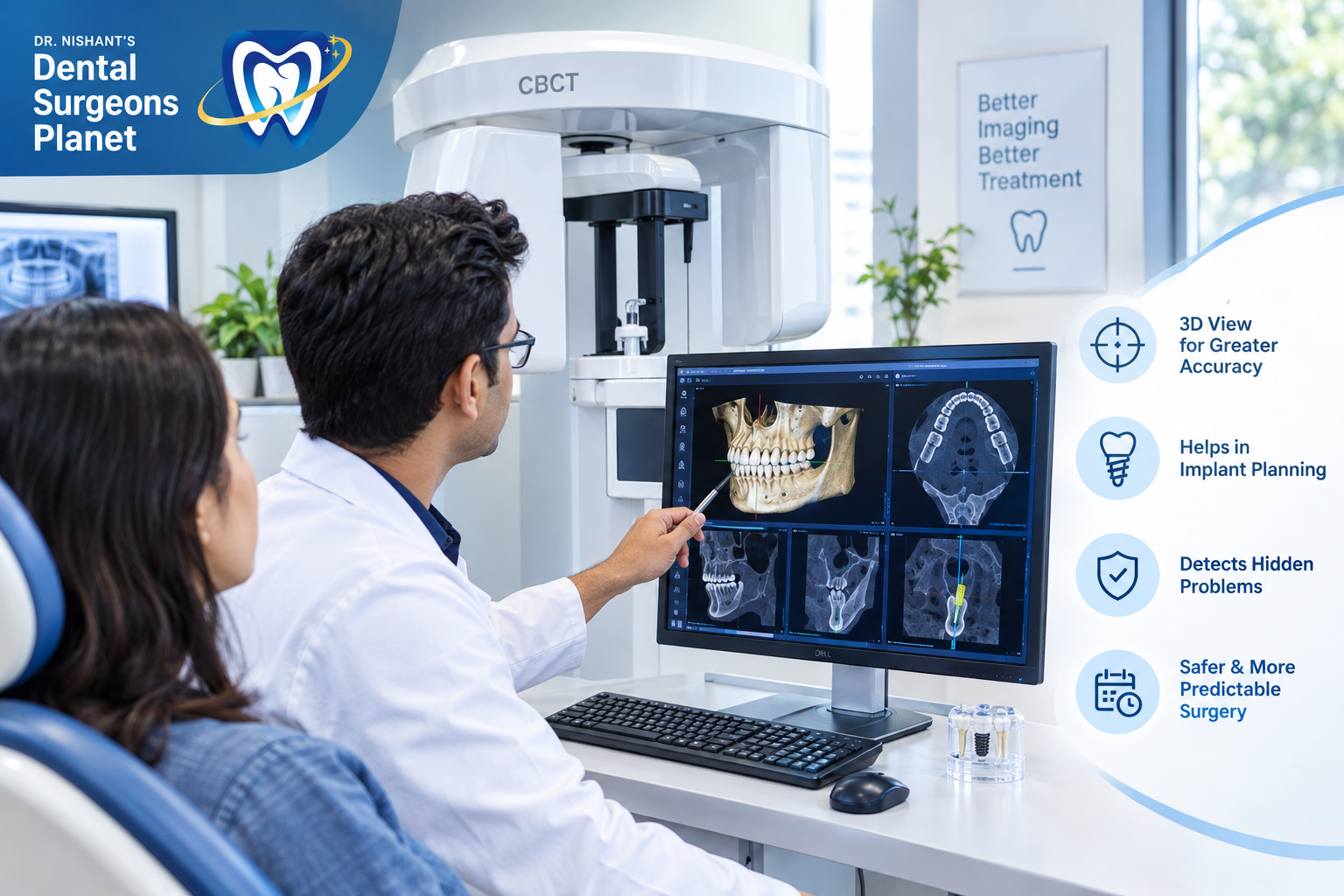
CBCT — Cone Beam Computed Tomography — is a specialised three-dimensional imaging technology used in dentistry that produces a detailed volumetric scan of the teeth, jawbone, nerves, sinuses, and surrounding structures in a single 10 to 40 second scan. The images it produces are fundamentally different from conventional dental X-rays in ways that directly determine surgical safety, implant success, and treatment outcomes.
Understanding what a CBCT scan shows — and what a conventional X-ray cannot — makes the reason your dentist is recommending it entirely clear.
What Is a CBCT Scan? The Simple Explanation
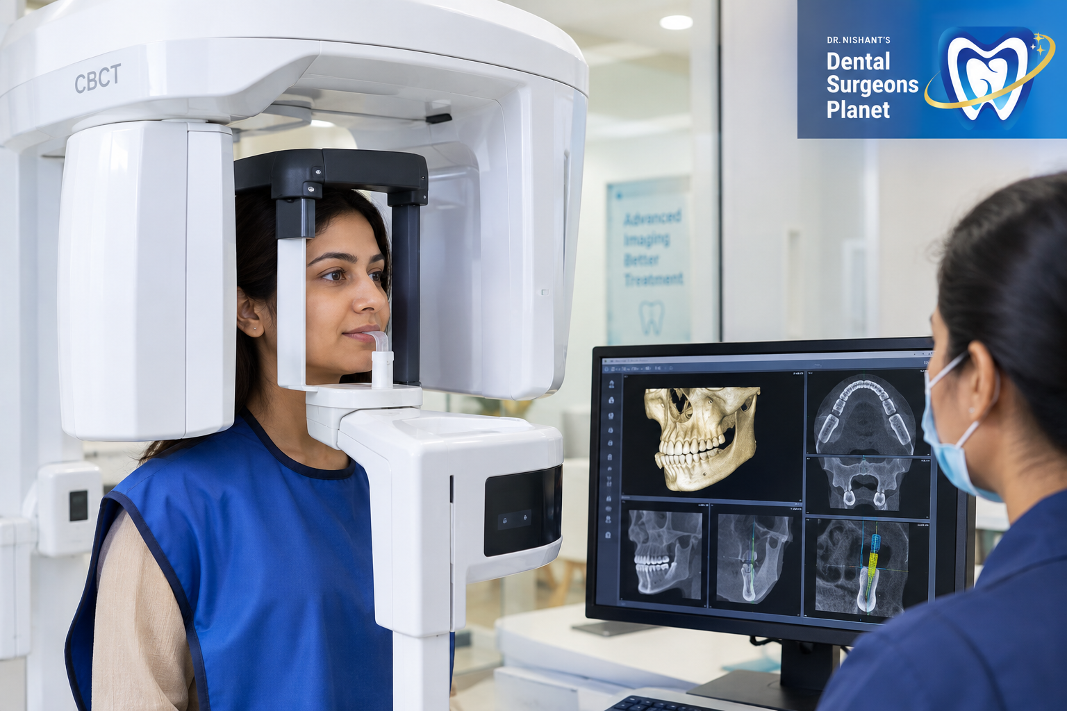
A CBCT scanner is a large machine — similar in appearance to a medical CT scanner but significantly smaller and designed specifically for dental and maxillofacial imaging. You stand or sit within the machine while a rotating arm moves around your head, capturing hundreds of two-dimensional X-ray images from different angles in a single rotation.
Sophisticated software then reconstructs these hundreds of images into a complete three-dimensional volumetric dataset of your jaw and surrounding structures. This dataset can be viewed as a three-dimensional model, or sliced in any direction — axial (from above), coronal (from the front), and sagittal (from the side) — to examine any specific anatomical area in complete detail.
The result is a precise, three-dimensional map of everything inside your jaw — the bone volume and density at every point, the exact position of every nerve canal, the location of anatomical structures like the sinus floor, and the relationship between all of these structures and the planned implant or surgical site.
This is information that a conventional two-dimensional X-ray fundamentally cannot provide — not because of image quality, but because of geometry. A flat image collapses a three-dimensional structure into two dimensions, making it impossible to measure depth, assess density across a volume, or understand the three-dimensional relationships between structures that surgical planning requires.
CBCT vs Conventional Dental X-Rays — The Critical Difference
Most patients have had dental X-rays — either small periapical films of individual teeth, or a panoramic OPG that shows the entire jaw in one image. Both are valuable diagnostic tools used daily at dental clinics across Powai Mumbai. But both share a fundamental limitation.
What conventional X-rays show
A periapical X-ray shows the crown and root of a tooth, the surrounding bone level, and the root canal anatomy in two dimensions. An OPG shows the complete dentition, jaw bones, sinuses, and temporomandibular joints in a single flat image.
Both are taken from a single angle and produce a single flat image. All three-dimensional information is collapsed and superimposed onto that single plane.
What this means practically for implant planning
When planning a dental implant in Powai Mumbai, your surgeon needs to know the width of the bone at the implant site — how much bone exists in the bucco-lingual dimension, the depth going from the front to the back of the jaw. This dimension is completely invisible on a conventional X-ray because the X-ray is taken from the front and collapses this depth dimension entirely.
Your surgeon also needs to know exactly where the inferior alveolar nerve — the major nerve running through the lower jaw — is positioned relative to the planned implant site. On an OPG, this nerve canal appears as a line in two dimensions. But the nerve does not run in a perfectly straight line in three dimensions — it curves, branches, and varies in depth in ways that only a CBCT reveals.
Placing an implant that inadvertently contacts or damages this nerve causes permanent numbness or altered sensation in the lower lip, chin, and teeth on that side — a complication that a CBCT scan identifies the risk of and allows the surgeon to plan around with complete precision.
What CBCT shows that X-rays cannot
Bone width and volume in all three dimensions — the most critical measurement for implant planning.
Bone density — the quality of bone at the implant site, which directly affects how the implant will integrate. Dense cortical bone integrates differently from soft cancellous bone, and the ratio between them at any given site is only assessable in three dimensions.
Precise nerve canal position in three dimensions — not just depth on a flat image but the complete three-dimensional course of the nerve through the jaw.
Sinus floor anatomy — the exact position of the maxillary sinus floor relative to planned upper jaw implant sites, determining whether sinus lifting is required and by how much.
Root positions of adjacent teeth — in three dimensions, allowing surgical access to be planned without risk of damaging neighbouring roots.
Pathology invisible on conventional X-rays — cysts, infections, and abnormalities that are hidden behind other structures on flat images but clearly visible in three-dimensional reconstruction.
When Is a CBCT Scan Required? — Clinical Indications
Not every dental appointment requires a CBCT scan. The additional cost and additional radiation — though modest relative to medical CT scans — are justified only when the three-dimensional information changes the treatment plan or significantly improves surgical safety compared to what conventional X-rays provide.
Here are the specific situations at Dental Surgeons Planet Powai where CBCT imaging is part of standard treatment planning:
Dental implant planning
This is the most common indication for CBCT in general dental practice. Before placing any implant, a CBCT scan at the planned implant site provides bone width measurements, bone density assessment, nerve canal location, and sinus anatomy — all essential for choosing the correct implant dimensions, planning the precise insertion angle, and determining whether bone grafting or sinus lifting is required before implant placement.
A dental implant placed without CBCT imaging in the lower jaw is placed with incomplete information about nerve canal position. A dental implant placed without CBCT in the upper jaw is placed with incomplete information about sinus floor position. Both represent avoidable surgical risks when CBCT is available and its cost is proportionate to the implant investment.
Oral and maxillofacial surgery
Surgical removal of impacted wisdom teeth — particularly lower third molars — frequently requires CBCT assessment when the OPG suggests close proximity between the impacted tooth roots and the inferior alveolar nerve canal. A CBCT confirms whether the roots are genuinely in contact with, wrapped around, or merely near the nerve canal — a distinction that determines whether standard surgical removal or a more specialised technique called coronectomy is the safer approach.
Jaw surgery — orthognathic surgery to correct skeletal jaw discrepancies — requires CBCT for complete three-dimensional surgical planning. The three-dimensional bone models generated from CBCT data allow virtual surgical simulation where the planned jaw movements are performed digitally before the actual surgery, and custom surgical guides are fabricated to translate the digital plan precisely to the operating theatre.
Cyst and tumour assessment requires CBCT to understand the full extent of the lesion — its boundaries in all dimensions, proximity to vital structures, and impact on surrounding bone — before surgical removal is planned.
Complex endodontic cases
Calcified root canals — where the canal space has narrowed significantly due to secondary dentine deposition — can be located with far greater accuracy using CBCT than conventional X-rays. Failed root canals where the cause of failure is unclear benefit from CBCT assessment — identifying missed canals, root fractures, or periapical pathology that flat X-rays cannot confirm. CBCT is also used for diagnosis of vertical root fractures where clinical examination and conventional radiographs are inconclusive.
Orthodontic planning for impacted teeth
Canine teeth are the most commonly impacted teeth after wisdom teeth. When an upper canine fails to erupt normally, CBCT reveals its precise three-dimensional position — how deeply buried, at what angle, and how close to adjacent roots — enabling precise surgical exposure planning and orthodontic traction direction to be determined before treatment begins.
Temporomandibular joint assessment
The TMJ — the joint connecting the lower jaw to the skull — has complex three-dimensional anatomy that conventional X-rays reveal only partially. CBCT provides detailed imaging of the joint surfaces, disc space, condylar morphology, and bone quality in patients with significant TMJ disorders being considered for surgical intervention.
Bone grafting planning
When significant bone loss has occurred following tooth extraction, advanced gum disease, or jaw pathology, CBCT quantifies the bone deficiency in three dimensions and guides graft volume planning before regenerative procedures or implant placement in grafted sites.
How Much Radiation Does a CBCT Scan Involve?
Radiation exposure is a legitimate concern for patients, and the honest answer involves context.
A conventional panoramic OPG delivers approximately 14 to 26 microsieverts of radiation. A full-mouth series of periapical X-rays delivers approximately 35 microsieverts.
A dental CBCT scan delivers between 40 and 200 microsieverts depending on the field of view — the size of the area being scanned. A small field of view scan focused on a single implant site delivers toward the lower end of this range. A large field of view full jaw scan delivers toward the higher end.
For context — natural background radiation from the environment delivers approximately 2,400 microsieverts per year to every person regardless of any dental imaging. A single transatlantic flight delivers approximately 50 to 100 microsieverts of cosmic radiation. A medical chest CT scan delivers approximately 7,000 microsieverts.
A dental CBCT scan at the doses used in dental practice represents a small fraction of annual background radiation exposure and is orders of magnitude below medical CT dosing. For adults undergoing dental implant surgery or complex oral surgery in Powai Mumbai — where the alternative is proceeding with incomplete anatomical information — the diagnostic benefit is entirely proportionate to the minimal radiation involved.
CBCT is used selectively, not routinely. It is recommended when the three-dimensional information changes the treatment plan or meaningfully improves safety. It is not used as a substitute for conventional X-rays for routine dental assessment.
The CBCT Scanning Experience — What Actually Happens
Patients who have never had a CBCT scan often imagine an experience similar to a hospital MRI or CT scanner — intimidating, enclosed, lengthy. The dental CBCT experience is considerably simpler.
Before the scan
You are asked to remove jewellery, glasses, and any removable dental appliances — anything metallic that would create artefact in the image. No special preparation, no fasting, no injection of contrast material is required for dental CBCT.
During the scan
You stand or sit in an open frame — nothing closes around you, there is no tunnel to pass through. Your head is positioned using chin and head rests to stabilise it during the rotation. You are asked to remain still and bite gently on a small bite guide that keeps your jaws in the correct position.
The rotating arm moves around your head once — taking 10 to 40 seconds depending on the machine and field of view selected. You hear a gentle clicking or whirring sound. You feel nothing. The scan is completely painless.
After the scan
The three-dimensional dataset is immediately available on the workstation. Your dentist or specialist reviews it on screen within minutes and can show you the three-dimensional reconstruction of your own jaw — explaining exactly what the scan shows in terms of bone volume, nerve position, and treatment planning implications in real time.
Total time in the scanning suite — 5 to 10 minutes including positioning and scan acquisition.
How CBCT Changes Implant Treatment Planning — A Specific Example
To make the clinical value of CBCT concrete, consider this specific scenario that occurs regularly at dental clinics in Powai Mumbai:
A patient presents for a single lower molar implant. An OPG is taken and shows adequate vertical bone height above the nerve canal — approximately 12mm. Based on the OPG alone, a 10mm implant appears safe with 2mm of margin from the nerve.
A CBCT is then taken. The three-dimensional data reveals two critical findings invisible on the OPG. First, the bone width at the implant site is only 5.5mm in the bucco-lingual dimension — insufficient for a standard 4.0mm diameter implant without encroaching on thin cortical bone walls. A narrower 3.5mm diameter implant must be selected or bone grafting performed first.
Second, the nerve canal has a buccal loop at the mesial aspect of the implant site — a curve toward the outer surface of the jaw that brings it 2mm closer to the implant path than the OPG suggested at that specific point.
With the OPG only — a standard diameter implant at planned length would have been placed with inadequate bone width and genuine nerve proximity risk. With the CBCT — a narrower implant is selected, the angulation is adjusted 3 degrees to avoid the nerve loop, and the patient has a successful implant procedure with zero complications.
This scenario is not unusual. It illustrates precisely why CBCT imaging before implant placement is standard of care rather than an optional upgrade at a clinic committed to genuine patient safety.
CBCT Scan Cost in Powai Mumbai
| Scan Type | Approximate Cost |
|---|---|
| Small field of view (single site) | ₹2,000 – ₹4,000 |
| Medium field of view (single jaw) | ₹3,000 – ₹6,000 |
| Large field of view (full jaw) | ₹5,000 – ₹10,000 |
| CBCT with implant planning software | ₹6,000 – ₹12,000 |
Some dental clinics in Powai Mumbai have in-house CBCT scanners. Others refer patients to dedicated dental radiology centres where the scan is performed and the DICOM data file sent to the treating dentist for planning. Both approaches are clinically equivalent — what matters is that the treating dentist reviews the three-dimensional data personally and integrates it into the treatment plan.
The cost of a CBCT scan represents a small fraction of the total implant or surgical treatment investment. In the context of a dental implant costing ₹25,000 to ₹60,000, spending ₹3,000 to ₹8,000 on imaging that makes the procedure safe and precisely planned is not an optional extra — it is a fundamental component of responsible treatment.
Questions to Ask Your Dentist About CBCT Before Treatment
If your dentist in Powai Mumbai has recommended a CBCT scan, these questions help you understand exactly why it is being recommended for your specific case:
What specific anatomical information does the CBCT provide that the OPG or periapical X-ray I already have does not show?
Will you review the CBCT personally and show me what it reveals about my jaw anatomy before finalising the treatment plan?
Does your clinic have an in-house CBCT scanner or will I need to attend a separate imaging centre?
How does the information from the CBCT change the implant dimensions, angulation, or surgical approach compared to what could be planned from conventional X-rays?
Is bone grafting or sinus lifting required based on the CBCT findings — and how would you know this without the scan?
A dentist who answers these questions clearly and specifically — rather than generically — is one who genuinely uses the CBCT data in clinical decision-making rather than simply recommending it as a routine additional charge.
FAQs
Q1. Is a CBCT scan the same as a regular dental X-ray?
No. A conventional dental X-ray produces a flat two-dimensional image from a single angle. A CBCT scan captures hundreds of images during a rotating scan and reconstructs them into a complete three-dimensional volumetric dataset. The information available from both is fundamentally different — CBCT provides bone width, density, nerve position, and three-dimensional anatomical relationships that flat X-rays cannot show.
Q2. Is a CBCT scan safe? How much radiation does it involve?
Yes, CBCT is safe for adults undergoing dental implant or surgical treatment. Dental CBCT delivers 40 to 200 microsieverts of radiation depending on the field of view — comparable to a few days of natural background radiation and significantly lower than a medical CT scan. It is recommended selectively when the diagnostic benefit justifies the exposure, not as a routine addition to every dental visit.
Q3. Do I always need a CBCT before a dental implant?
At a clinic committed to safe, precisely planned implant surgery — yes. The bone width, density, and nerve canal position information that CBCT provides is essential for choosing the correct implant dimensions, planning the insertion angle, and confirming that grafting or sinus lifting is not required. Implants placed without this information in the lower jaw carry avoidable nerve proximity risk.
Q4. How long does a CBCT scan take at a dental clinic in Powai Mumbai?
The scan acquisition itself takes 10 to 40 seconds. Total time in the scanning suite including positioning is 5 to 10 minutes. No preparation or recovery time is required — you leave immediately after the scan.
Q5. Can children have CBCT scans?
CBCT is used in children for specific clinical indications — impacted teeth, orthodontic anomalies, and jaw pathology — where the diagnostic benefit justifies the exposure. As with adults, it is not used routinely but selectively when three-dimensional information is clinically necessary. Paediatric doses are lower than adult doses through appropriate machine settings and small field of view selection.
Q6. What is the difference between a dental CBCT and a hospital CT scan?
A hospital CT scanner uses a fan-shaped X-ray beam and produces cross-sectional images at higher resolution and significantly higher radiation doses than dental CBCT. Dental CBCT uses a cone-shaped beam, is specifically designed for dental and maxillofacial anatomy, delivers far lower radiation, and produces images optimised for bone and dental structure assessment rather than soft tissue contrast. They are different technologies designed for different diagnostic purposes.
Conclusion
A CBCT scan is not an optional imaging upgrade or an additional revenue item at a dental clinic in Powai Mumbai that recommends it responsibly. It is a fundamentally different class of diagnostic information — three-dimensional, volumetric, and precise — that changes treatment planning decisions, improves surgical safety, and reduces complication risk in ways that conventional X-rays simply cannot replicate.
For dental implants — understanding bone width, density, and nerve canal position in three dimensions before placing a titanium screw into the jaw is not excessive caution. It is the minimum information a surgeon needs to place that implant safely and in the correct position for long-term stability.
For oral surgery — knowing the three-dimensional relationship between an impacted tooth and the nerve canal before making a surgical incision is what separates a predictable, complication-free procedure from one with avoidable risk.
If your dentist in Powai Mumbai has recommended a CBCT scan before your implant or surgical treatment — they are recommending it because the information it provides directly affects how safely and precisely your treatment can be performed. That recommendation reflects clinical responsibility, not unnecessary expense.
Visit Dental Surgeons Planet in Powai Mumbai for dental implant and surgical treatment planned with complete CBCT imaging — because treatment planned with complete information is treatment planned safely.
Precise imaging. Precise planning. Precise results. Book your implant consultation in Powai Mumbai today.